ex vivo expanded endothelial progenitor cells
Cell Culture.
1. Total hPBMCs were isolated from blood of human volunteers by density gradient centrifugation.
2. Cells were plated on culture dishes coated with human fibronectin and maintained in EC basal medium-2 (EBM-2).
3. The media was supplemented with EGM-2 containing 5% FBS, human VEGF-1, human fibroblast growth factor-2 (FGF-2), human epidermal growth factor (EGF), insulin-like growth factor-1 (IGF-1), and ascorbic acid.
4. After 4 days in culture, non-adherent cells were removed by washing with PBS, new media was applied, and the culture was maintained through days 7–10.
5. Human umbilical vein ECs were prepared, and human microvascular ECs (HMVECs) from adult dermis were used.
Cellular Staining.
1. Fluorescent chemical detection of EPCs was performed on attached hPBMCs after 7 days in culture.
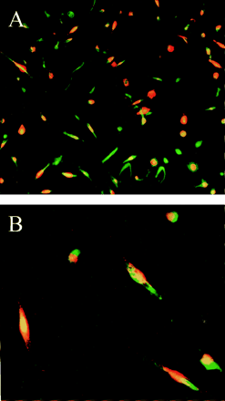
2. Direct fluorescent staining was used to detect dual binding of FITC-labeled Ulex europaeus agglutinin-1 and 1,1′-dioctadecyl-3,3,3′,3′-tetramethylindocarbocyanine (DiI)-labeled acetylated low density lipoprotein.
3. Cells were first incubated with acLDL at 37°C and later fixed with 1% paraformaldehyde for 10 min.
4. After washes, the cells were reacted with UEA-1 (10 μg/ml) for 1 h. After the staining, samples were viewed with an inverted fluorescent microscope.
5. Cells demonstrating double-positive fluorescence were identified as differentiating EPCs.
6. Cultured human umbilical vein ECs and NIH 3T3 cells served as positive and negative controls, respectively.
Fluorescence-Activated Cell Sorting.
1. Fluorescence-activated cell sorting (FACS) detection of EPCs was performed on attached hPBMCs after 7 days in culture.
2. Mononuclear cells were detached with the minimal use of trypsin and/or PBS with 1 mM EDTA followed by repeated gentle flushing through a pipette tip.
3. Cells (2 × 10⁵) were incubated for 30 min at 4°C with the monoclonal antibodies against kinase insert domain-containing receptor (KDR), recognizing the extracellular domain, and against human vascular endothelium (VE)-cadherin, the biotinylated hCD62E (E-selectin), the FITC-conjugated hCD3), hCD51/61 (αvβ3), hCD86, hCD83, hCD68, and the phycoerythrin-conjugated hCD31, hCD34, hCD14, and hCD19.
4. Isotype-identical antibodies served as controls.
5. For analysis of KDR and VE-cadherin, the cells were further incubated with a biotinylated anti-mouse IgG (H + L) antibody made in horse and with FITC-conjugated streptavidin.
6. After treatment, the cells were fixed in 1% paraformaldehyde.
7. Quantitative FACS was performed on a FACStar flow cytometer.
8. Histograms of cell number vs. logarithmic fluorescence intensity were recorded for 10,000–20,000 cells per sample.

In vitro differentiation of peripheral mononuclear cell subpopulation into EPCs. Endocytosis of acLDL (red fluorescence) and binding to UEA (green fluorescence) identified EPCs. (A) ×10 magnification; (B) ×40 magnification.
Reference
1. Asahara T, Murohara T, Sullivan A, Silver M, van der Zee R, Li T, Witzenbichler B, Schatteman G, Isner J M (1997) Science 275:965–967.
2. Brogi E, Schatteman G, Wu T, Kim E A, Varticovski L, Keyt B, Isner J M (1996) J Clin Invest 97:469–476.


